Phospholipid encapsulation enables enhanced ultrasound contrast and localized delivery of therapeutic gases following hypoxic-ischemic injury.
Problem:
Noble gases such as xenon (Xe), Argon (Ar) and helium (He) are known to provide cardioprotective and neuroprotective effects in hypoxic-ischemic injuries, traumatic brain injury and post-cardiac arrest. However, these gases are typically delivered systemically through inhalation which reduces their local therapeutic effects. Other delivery methods involve encapsulating the gases within liposomes injected into the carotid artery, then released locally through ultrasound exposure. Though this approach solves the local delivery issue, it is associated with potential complications due to carotid insertion, and shows poor ultrasound contrast. Improvement is needed to use noble gases as theranostic agents.
Solution:
Dr. Lee’s team at the Soft Materials Research and Technology (SMART) Lab developed an alternative localized delivery method using a mix of lipopolymers and phospholipids to generate noble gas microbubbles (NGMBs) ranging between 1 and 10 µm that are stable under physiological conditions and show high contrast when exposed to ultrasound.
Technology:
Disclosed herein is a process to produce physiologically stable NGMBs that are highly echogenic and amenable to localized delivery through retro-orbital injection. The NGMBs are produced through sonication and centrifugation with pure Xe, Ar or He in the presence of long chain phospholipids (DSPC) and a high molecular weight lipopolymer (DSPE-PEG5000). The inventors injected freshly prepared Xe-MBs intravenously behind the orbit of a mouse’s eye and succeeded to obtain ultrasound images of the heart and kidneys within seconds (see Image).
Advantages:
- 10 to 100-fold increased gas payload compared to liposomes
- Does not require perfluorocarbon additive as stabilizers
- Stable for up to 5 days after production
- Highly echogenic for at least 3 min even in the presence of serum proteins
- Avoids complications associated to carotid insertion
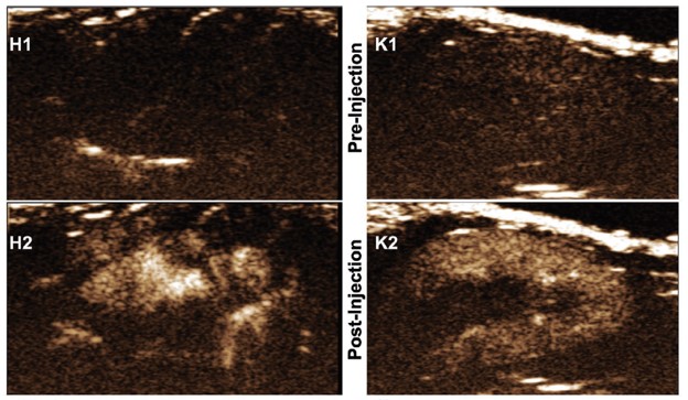
In vivo signal from the left ventricle of the heart (HI-H2) and kidney (K1-K2) of a mouse obtained by ultrasound following retro-orbital injection of 0.03 mL xenon microbubbles (XeMBs). Panels H1 and K1 show representative still frames from before injection, as panels H2 and K2 show representative still frames a few seconds after injection.
Stage of Development:
- Preclinical Discovery
- In vivo proof-of-concept
Case ID:
20-9168-TpNCS
Web Published:
6/16/2026
Patent Information:
| App Type |
Country |
Serial No. |
Patent No. |
File Date |
Issued Date |
Expire Date |