Targeted delivery of an anti-inflammatory cytokine to prevent heart failure after myocardial infarction.
Problem:
Heart failure (HF) is one of the leading causes of death worldwide and is expected to affect more than eight million people in the US by 2030. Myocardial infarctions (MIs) are the leading cause of HF due to excessive inflammation caused by MIs. Interleukin-4 (IL-4) is a cytokine that reduces inflammation and can potentially mitigate HF after MI, but systemic delivery can lead to off-target toxicity in surrounding tissues. Therefore, there is a need for therapeutics that sequester IL-4 to infarction sites.
Solution:
The inventors developed a protein complex called LMJ2.5I-IL4 that delivers IL-4 to myocardial infarction sites with minimal off-target effects. Targeted delivery of IL-4 increases local inflammation and efferocytosis by immune cells which reduces infarct size, reduces fibrosis, improves heart function, and prevents HF.
Technology:
The inventors fused IL-4 to LMJ2.5I, a custom nanobody that targets a fibronectin variant expressed in infarcted tissue. Using a gold-standard mouse model of MI, the inventors demonstrated that their technology delivers IL-4 to infarctions with little off-target effects. This high-precision delivery of IL-4 polarizes local immune cells towards protecting against HF.
Advantages:
- Provides significantly increased delivery of therapeutic to infarcts in a coronary artery ligation (LAD) mouse model of MI
- Improves left ventricular ejection fraction (45% versus 29% in control group) in mouse model of MI
- Decreases fibrosis (33% relative to control group) in mouse model of MI
- Increases reparative monocytes and macrophages at the infarction site in a mouse model of MI
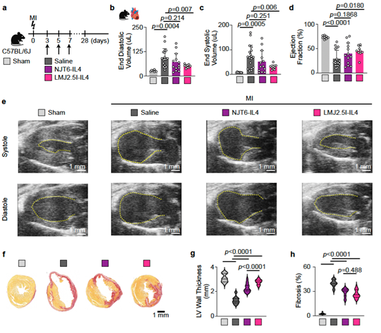
Fn-EIIIB targeted IL-4 immunocytokine improves long-term cardiac function after MI. a., Experimental outline. C57BL/6 mice were subjected to permanent coronary artery ligation (MI) or sham surgery and treated with either saline, NJT6-IL4, or LMJ2.5I-IL4 on days 3, 5, and 7 after surgery (n = 9-18 mice per treatment group after MI, n = 8 mice for Sham). b.-d., (b) Left ventricular end-diastolic volume, (c) end-systolic volume, and (d) ejection fraction on day twenty-eight after surgery. Data, shown as mean+/- S.D., were analyzed by one-way ANOVA and Tukey’s post hoc test. e., Representative parasternal long axis views of the left ventricles (outlined) in systole and diastole on day twenty-eight. Scale bar is 1 mm. f., Representative Picrosirius Red stained cardiac cross sections on day twenty-eight. Scale bar is 1 mm. g., Quantification of left ventricular wall thickness (n = 3 mice per group, 6-7 cross sections per mouse were analyzed). Data, shown as mean +/- S.D., were analyzed by one-way ANOVA and Tukey’s post hoc test. h., Quantification of fibrosis (n = 3 mice per group, 6-7 cross sections per mouse were analyzed). Data were analyzed by one-way ANOVA and Tukey’s post hoc test.
Stage of Development:
- Target Identified
- Preclinical Discovery
- IND Enabling Studies